Introduction
Back pain is a very common symptom in clinical practice, and physicians will need to face its initial management in any patient.[1] Although vertebral osteomyelitis should be included in the differential diagnosis of back pain, it is much less frequent than other spinal diseases and for this reason often recognized and treated too late.[1] Spondylitis, a osteomyelitis of the spinal column is defined as infection of the vertebral bodies, starting at the endplate, with secondary involvement of the intervertebral disc.[2] It can be of pyogenic, granulomatous, and parasitic etiology.[3]We present this case because typical symptoms of vertebral osteomyelitis were absent and the diagnosis was challenging.
Clinical Case
A 81 year-old man presented with an one year history of lumbago. He was a frequent consumer of unpasteurized dairy products and had no history of travel outside Europe. Thoracic computerized tomography [CT] and pulmonary bronchofibroscopy had been performed eleven years earlier because of hemoptysis, cough and a pulmonary lesion. Bronchoalveolar lavage culture revealed Mycobacterium tuberculosis [MT]. The patient was lost to follow-up and as he had no more symptoms, no treatment was initiated. Others major pathologies of his background were hypertension, diabetes, dyslipidemia and ischemic and hypertensive heart disease.
The patient was diagnosed with lumbar strain and medicated with analgesics. As a mechanical lumbar pain persisted, acupuncture was performed and the patient suffered a third degree burn. He had no history of fever, weight loss or anorexia. Also, there was no history of traumatic injury. On physical examination, no abnormalities were noted, including neurological deficits.Given the persistence and worsening of the lumbar pain, magnetic resonance imaging [MRI] was performed, revealing marked bone destruction of L4 and L5 and adjacent areas sclerosis and edema in the bone marrow, edema in areas of the body L3 and collapse and reduction of amplitude of intersomatic space of L4 and L5. Indentation of L4 and L5 wall was conditioning reduction of vertebral canal amplitude and infectious componentsin epidural canal in L3-L4, L4 -L5 and L5-S1 conditioned compression of the thecal sac. There were also a paraspinal left side infectious collection in the psoas muscle in continuity with the epidural collection of L3 -L4 with 36x21mm.
The patient was then hospitalized in our department to pursue investigation. As the patient had multiple predisposing factors for different pathogens, etiological differential diagnosis included MT, Brucella and Gram-positive cocci. As the patient was stable, no empirical antibiotics were initiated.On admission, the erythrocyte serum sedimentation rate was normal [22mm/h], Human Immunodeficiency Virus was negative; and all other blood tests were unremarkable.Transthoracic echocardiogram was normal and blood cultures were negative. Bone marrow and sputum culture were negative.CT-guided paravertebral abscess aspiration was performed. Cultures were negative but DNA amplification by the polymerase chain reaction was positive for Mycobacterium tuberculosis, making a final diagnosis of Potts disease.
Treatment with isoniazid, rifampicin, pyrazinamide and ethambutol was initiated.
After initiation of the treatment and evaluation of its tolerability, the patient was discharged. After two months of quadruple therapy, he maintained isoniazid and rifampicin only during the next 7 months. The patient maintained follow-up in our clinic and showed both clinical and radiological amelioration. As he had no neurologic symptoms, neurosurgery was deemed unnecessary.
Discussion
Potts disease remains a rare cause of back pain and its diagnostic workup is challenging. The absence of constitutional symptoms in a high percentage of cases explains why many physicians fail to consider infection as the cause of back pain.[1] A high index of suspicion is needed for the diagnosis of Potts disease especially given the insidious onset of symptoms and reports of a long period between onset of symptoms and diagnosis of disease.
Although thoracic segment and the thoracolumbar hinge represent the most frequent localization of tuberculous spondylodiscitis [TS], in our case only the lumbar segment was affected, making diagnosis more difficult. [4] TS are frequently complicated by formation of large paravertebral abscesses, which have been reported in 20-90% of cases.[4]
Early diagnosis and therapy are important to prevent function loss and morbidity and to achieve normalization of the patient´s daily activities. For this reason, specific antibiotic therapy is one of the keystones of spondylodiscitis treatment and this requires specific identification of the pathogen and determination of its sensitivity to antibiotics.[2, 5]
The basic principles that underlie the treatment of pulmonary tuberculosis also apply to extra-pulmonary forms of the disease. Although relatively few studies have examined treatment of extra-pulmonary tuberculosis, increasing evidence suggests that 6- to 9-month regimens that include isoniazid and rifampicin are effective.[6, 7]
Possible indications for surgical intervention are: presence of neurological deficits caused by spinal cord compression, spinal deformity with instability, severe or progressive kyphosis, no response or failure of antituberculous therapy, large spinal abscesses and non-diagnostic biopsies, even if repeated.[4]
In general, younger age and early diagnosis have been reported as factors associated with a good outcome, whereas presence of paraplegia at presentation has been recognized as the most negative prognostic factor.[4]
Figura I
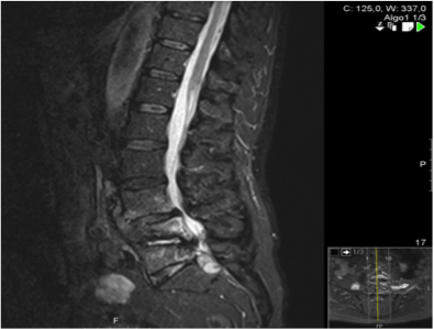
Ressonância Magnética da coluna lombar, ponderação em T2
Figura II
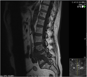
Ressonância Magnética da coluna lombar, ponderação em T1
BIBLIOGRAFIA
1. Colmenero J, Ruiz-Mesa J, Sanjuan-Jimenez R, Sobrino B, Morata P. Establishing the diagnosis of tuberculous vertebral osteomyelitis. Eur Spine J. 2013;22[4]:579-86.
2. Sobottke R, Seifert H, Fatkenheuer G, Schmidt M, Gossmann A, Eysel P. Current diagnosis and treatment of spondylodiscitis. Deutsches Arzteblatt international. 2008;105[10]:181-7.
3. Gouliouris T, Aliyu SH, Brown NM. Spondylodiscitis: update on diagnosis and management. The Journal of antimicrobial chemotherapy. 2010;65 Suppl 3:iii11-24.
4. Trecarichi EM, Di Meco E, Mazzotta V, Fantoni M. Tuberculous spondylodiscitis: epidemiology, clinical features, treatment, and outcome. European review for medical and pharmacological sciences. 2012;16 Suppl 2:58-72.
5. Capelo J, Carragoso A, Albuquerque C, Mocho ML, Canto-Moreira N. [Infectious spondylodiscitis: a study of forty-one cases]. Acta reumatologica portuguesa. 2007;32[3]:255-62.
6. Blumberg HM, Burman WJ, Chaisson RE, Daley CL, Etkind SC, Friedman LN, et al. American Thoracic Society/Centers for Disease Control and Prevention/Infectious Diseases Society of America: treatment of tuberculosis. American journal of respiratory and critical care medicine. 2003;167[4]:603-62.
7. Blumberg HM, Leonard MK, Jr., Jasmer RM. Update on the treatment of tuberculosis and latent tuberculosis infection. JAMA : the journal of the American Medical Association. 2005;293[22]:2776-84.

