Due to the exuberance of the cutaneous lesions, we report the case of a 43-year-old woman, admitted to the emergency department with an itchy rash with one week of evolution. She started about 2 weeks before taking daily oral terbinafine to treat persistent onychomycosis of the toe nails. There was no family history of similar illness. No previous illnesses or usual medication. There was no history of fever or preceding upper respiratory illness. She was not on any other medication simultaneously and never had a similar rash in the past. There was no history of previous drug allergy. Upon observation, she presented erythematous rash on face and target lesions in the arms and legs (Fig. 1 and 2). No lesions of the mucous membranes, hands or feet. Investigation only revealed a slight increase in c-reactive protein. A skin biopsy was performed, which revealed the presence of spongiform pustules, capillary dilation and peri-vascular lymphohistiocytic infiltrate, with evident foci of neutrophil exocytosis, data compatible with erythema multiforme. After exclusion other causes, terbinafine was withdrawn. The patient was given supportive management in the form of antihistamines and steroids. The rash began to resolve quickly, and the patient was discharged from hospital after 8 days.
Erythema multiforme (EM) is an acute and sometimes recurring skin inflammatory condition.1 In most cases, it is secondary to infections (mainly herpes simplex virus, adenovirus, enterovirus and Mycoplasma pneumonia) and, less frequently, to drugs. Drug-associated EM is reported in 10-50% of cases.2 Erythema multiforme have been associated with the use of oral antifungals in a number of case reports and retrospective series of cases.3,4 Genetic susceptibility can be a predisposing factor in some patients with EM.2 EM is an uncommon mucocutaneous disorder, characterized by targetoid lesions on the skin,5 with a symmetrical distribution over the dorsal surfaces of the extensor members and minimal involvement of the mucous membranes.6 Occasionally EM may involve the mouth alone.2 EM spectra have been classified mainly as minor and major forms: EM minor typically affects only one mucosa (generally the oral mucosa) and may be associated with symmetrical target skin lesions on the extremities; EM major typically involves two or more mucous membranes with more variable skin involvement.2 The mechanism of tissue damage in EM seems to vary in infection-induced EM and drug-induced EM. Infection-induced EM appear to be a result of the delayed type hypersensitivity reaction, with production of IFN-γ, chemokines and cytocines, recruitment of cytotoxic T cells and epitelial damage. In drug-induced EM, reactive metabolites of the particular drug induce the disease and keratinocyte apoptosis is induced by TNF-α released from keratinocytes, macrophages and monocytes, leading to tissue damage.2 EM is usually self-limiting, resolving in weeks without significant sequelae.
Figura I
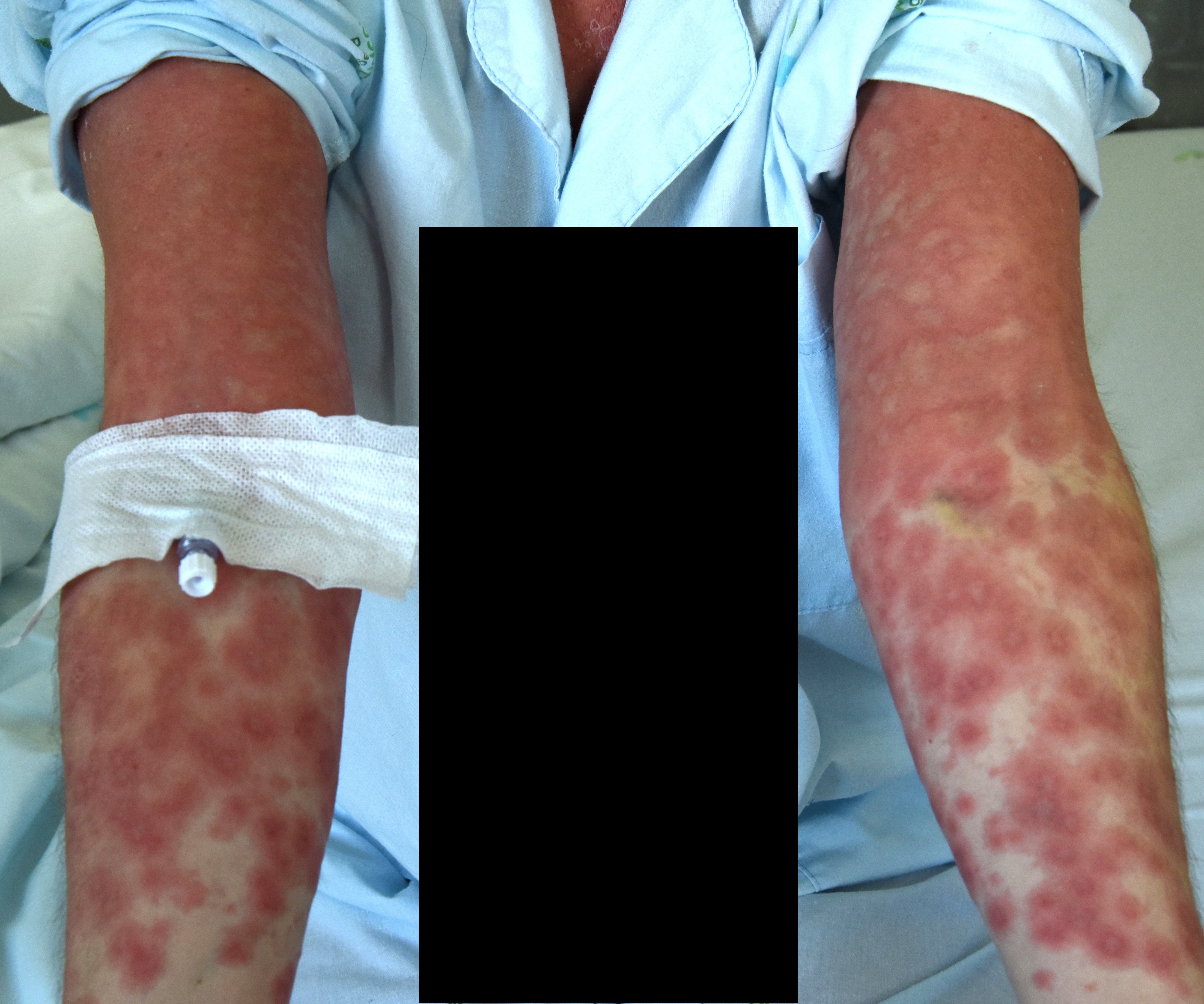
Figure 1 Erythematous rash in patients arms, symmetrically arranged, with the classical target lesion appearance.
Figura II

Figure 2 Pink-red blotches with the classical target lesion appearance (pink-red ring around a pale center) in patients arms.
BIBLIOGRAFIA
[1] Sokumbi O, Wetter DA. Clinical features, diagnosis, and treatment of erythema multiforme: a review for the practicing dermatologist. Int J Dermatol. 2012 Aug. 51(8):889-902.
[2] Krishnankutty K N, Chaudhuri k, Ashok L. Erythema multiforme: a case series and review of literature. Open Access J Trans Med Res. 2018; 2(4):124-130.
[3] Castellsague J, García-Rodríguez LA, Duque A, Pérez S. Risk of serious skin disorders among users of oral antifungals: a population-based study. BMC Dermatol. 2002 Nov 28;2:14.
[4] George A, Bhatia A, Kanish B, Williams A. Terbinafine induced pityriasis rosea-like eruption.Indian J Pharmacol 2015;47:680-1
[5] Atzori L, Pau M, Aste M. Erythema multiforme ID reaction in atypical dermatophytosis: a case report. J European Academy of Dermatology and Venereology. 2003 Nov. 17(6):699-701
[6] Gupta, Lynde, Lauzon, et al. Cutaneous adverse effects associated with terbinafine therapy: 10 case reports and a review of the literature. British J Dermatol. 1998 March. 138(3):529-32.

