Case report
Patient showed widespread erythematous rash and cutaneous nodules around arms and abdomen over a period of 2 months characterized by non tender, non pruritic papules and erythemato-violaceous nodules, 0.5 cm in diameter (fig 1-2). No palpable peripheral lymphadenopathy nor history of reported night sweats or weight loss. Peripheral blood smear revealed 76% neutrophilia, 2% myelocytes and 8% rods. Punch biopsy of the lesions was taken and submitted for pathologic evaluation. The histopathological study showed skin infiltration by atypical cells in the epidermis, dermis and subcutaneous tissue with a perivascular pattern. Immunohistochemistry showed diffuse positivity for CD20, CD79α and Bcl2 neoplastic cells, with heterogeneous and focal positivity for ACL, MUM1, Bcl6, PAX5 and CD23. Biopsy was suggestive lymphoid B cell neoplasia involvement. Systemic disease was excluded.
Discussion
LC lesions are rare and diagnosis may be difficult. Lesions may take different appearances and are highly variable in the presentation. Usually presents as a manifestation of previously diagnosed acute or chronic leukemias, lymphoid malignancies and myelodysplastic syndromes but in rare cases, LC may precede the development of systemic disease1-2. It also makes a differential diagnosis with primary cutaneous lymphoma, which must be excluded3.
Histophatologic examination of the skin lesions shows infiltration of dermis and hypodermis by blast cells with perivascular and perianexial predominance. In myeloid leukemia the epidermis is respected, however, in lymphoid leukemias diffuse infiltrates are observed and leukemic cells have epidermotropism, as in our patient4.
Literature on aleukemic leukemia cutis is limited, but because diagnosis of leukemia cutis portends poor prognosis in acute leukemia, the treatment should be directed at eradicating the leukemic clone by using aggressive systemic chemotherapy and stem cell transplant possibly in first remission, however due to the advanced age of our patient and low physiological reserve, the patient started on Radiotherapy, because is beneficial in widespread skin involvement
Figura I
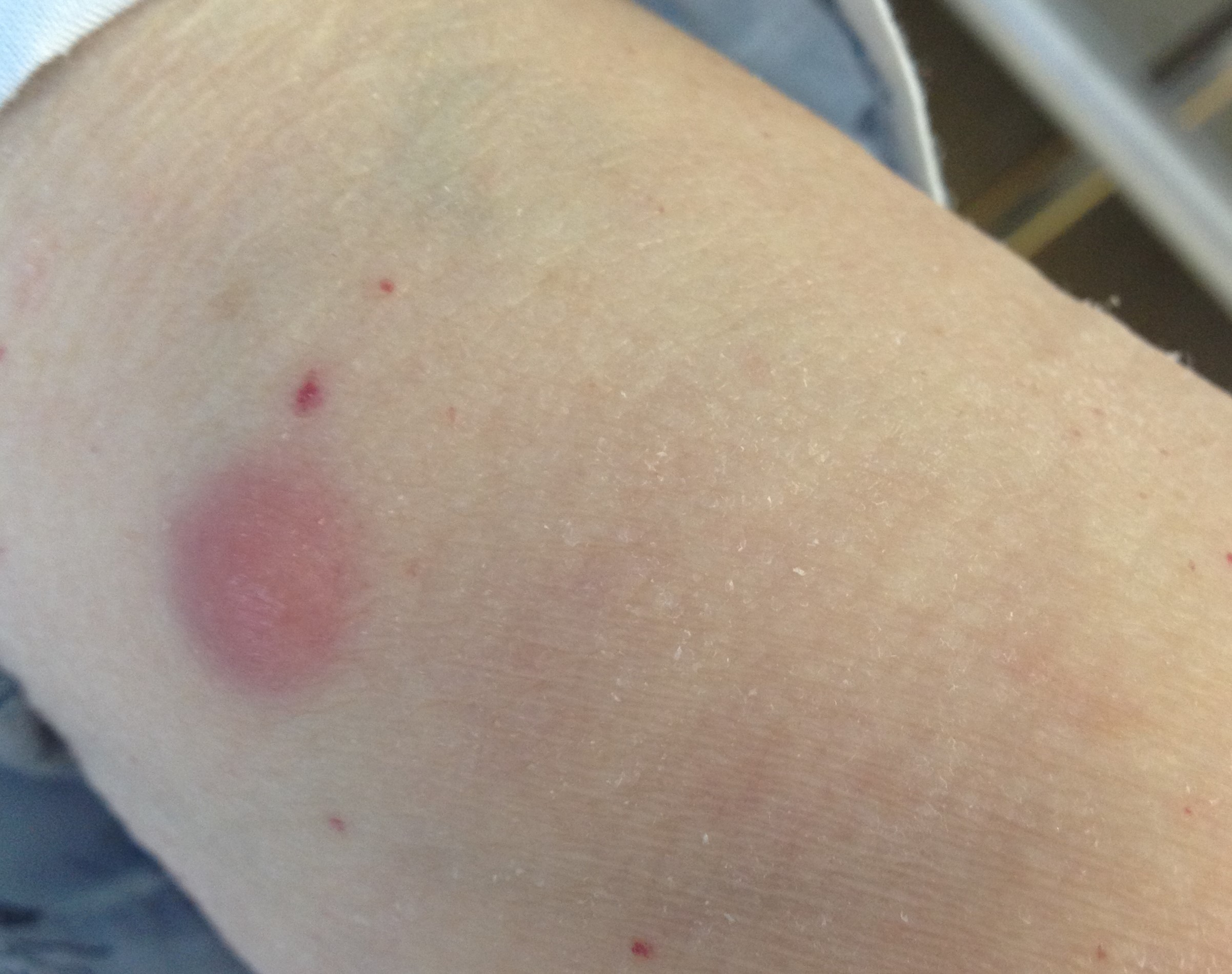
Lesion around arm
Figura II
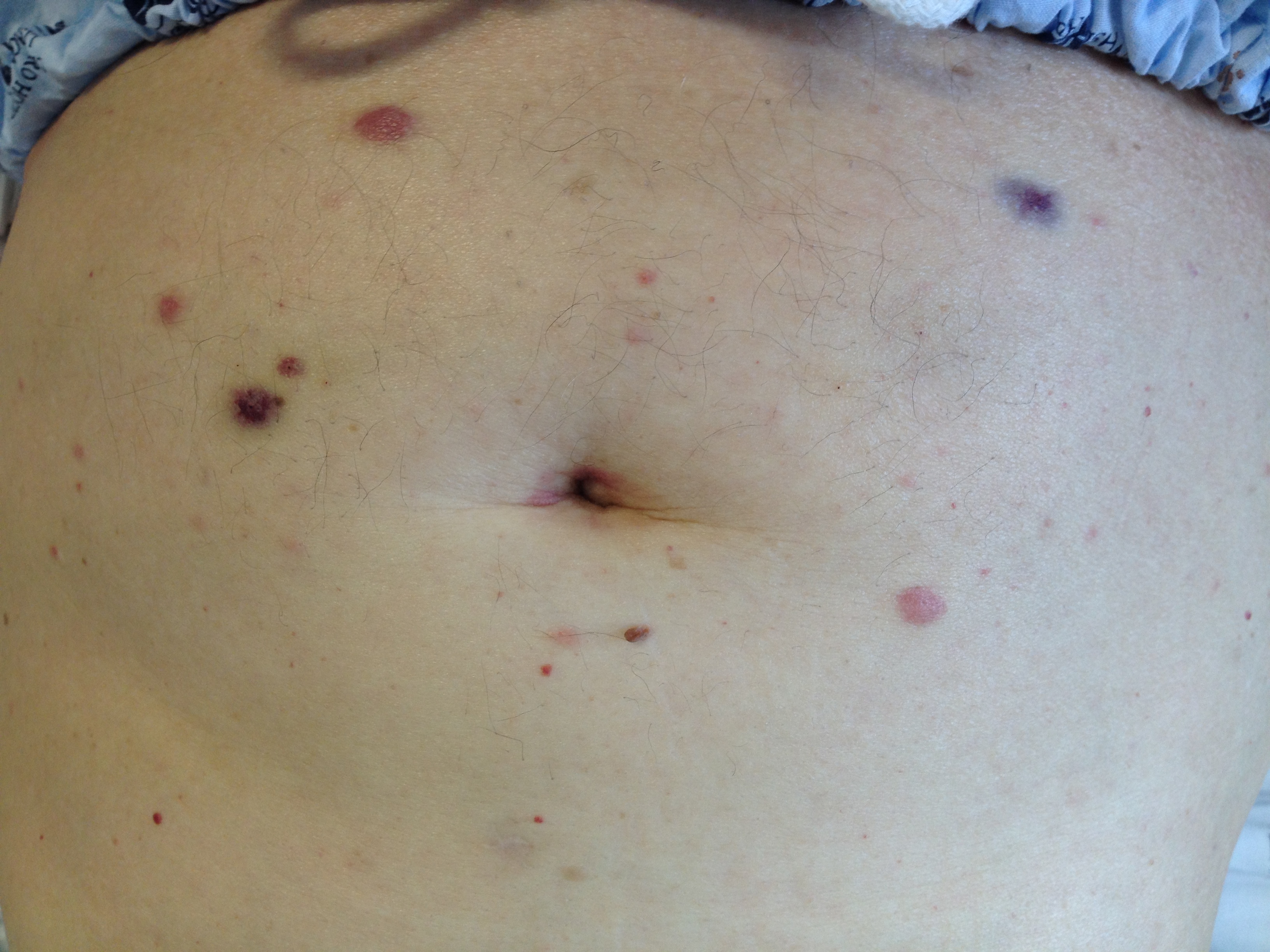
lesions around abdomen
BIBLIOGRAFIA
1. Rao AG, Danturty I. Leukemia cutis.Indian J Dermatol. 2012;57(6):504. doi:10.4103/0019-5154.103086
2.Kang YS, Kim HS, Park HJ, et al. Clinical characteristics of 75 patients with leukemia cutis.J Korean Med Sci. 2013;28(4):614619. doi:10.3346/jkms.2013.28.4.614
3. Ngan AV, Writer S, Menezes S De, Trainee BP, Health A. Cutaneous B-cell lymphoma Primary cutaneous follicle centre lymphoma. 2021;(November 2016):16.
4. Haidari W, Strowd LC. Clinical characterization of leukemia cutis presentation. Cutis. 2019 Dec;104(6):326-330;E3.

