INTRODUCTION
Hyponatremia (sodium < 135 mEq/L) poses a great diagnostic challenge.1,2 Clinically, its course is marked by the timing of installation, being acute hyponatremia (developed in less than 48 hours) the most expressive one. When considering chronic hyponatremia (elevated natremia for longer than 48h), clinical features may be subtle, delaying its diagnosis and its correction needs to occur at a slower rate, posing some difficulties in patients approach. Syndrome of Inappropriate Antidiuretic Hormone Secretion (SIADH) is one of the most frequent aetiologies and is characterized either by inappropriate secretion of antidiuretic hormone (ADH) from its normal hypothalamic source or by ectopic production. Both ways lead to euvolemic hyponatremia, serum hypoosmolarity, elevated urine osmolality (>100 mOsm/kg) and urinary sodium (> 20 mEq/L). SIADH englobes five broad etiological categories (table 1): nervous system disorders, neoplasia/paraneoplastic syndromes, pulmonary diseases, drug induced or other miscellaneous aetiologies.1,2 Amongst the neurological causes, hydrocephalus may be one of the causal factors, although considered a rare one, making high clinical suspicion fundamental for its diagnosis and appropriate treatment.3,4 We present a case of SIADH due to secondary normal pressure hydrocephalus (NPH) in the presence of aqueductal membrane stenosis caused by a membrane.
CLINICAL CASE
We present the case of a 67-year-old female patient, with medical history of hypertension; aortic stenosis with previous implantation of prosthetic valve and coronary ischaemic disease, without heart failure; and active smoker (20 pack-years). She was being treated with several medications none of them a diuretic (losartan 100mg q.d., amlodipine 5mg q.d, nebivolol 5mg q.d. and acetylsalicylic acid 100mg q.d.). The patient presented four times to the emergency department (ED) with 1 yearlong complaints of urinary urgency. In the first three times, she was discharged on antibiotics, admitting urinary tract infection, but without microbiological confirmation. In the fourth time, urine tests showed no leukocyturia nor pyuria, nitrites were negative and urine cultures were sterile and blood tests showed hyponatremia (123 mEq/L) without worsening of kidney function nor other electrolyte abnormalities. While on the ED, the patient started fluid repletion with sodium chloride 0.9% and was admitted to the internal medicine ward for surveillance and etiological study. Upon further investigation, the patient reported gait disturbance (lasting 4 years) as well as memory loss episodes. On examination the patient was conscious, alert, oriented in time, place and person; her speech was normal, as well as calculus and attention; she was afebrile, normotensive, normocardic and euvolemic (jugular venous pressure was normal); and there was no peripheral oedema nor pulmonary rales). Neurological examination was normal except for a broad-based gait. On the ward, due to lack of improvement of the sodium levels, fluid replacement was stopped, and hyponatremia workup was done after a 24-hour period window: serum osmolality 275 mOsmol/kg (reference value 275-300 mOsmol/kg), urine osmolality 182 mOsmol/kg (reference value 50-1200 mOsmol/kg), urine sodium 31mEq/L; normal thyroid function and cortisol levels. Hyponatremia resistant to fluid administration coupled with high urine osmolality pointed towards the diagnosis of SIADH. A presumed SIADH diagnosis was made, and the patient started a fluid restriction diet (less than 1 litre per day), consequently normalizing natremia levels (reference value 136-145 mEq/L). A diagnostic full body computed tomography (CT) scan was performed to rule out ectopic sources of ADH production. The cranioencephalic CT showed supratentorial ventriculomegaly, in particular of the atrium and occipital horns of the lateral ventricles and third ventricle, with no unmistakable signs of trans-perpendicular oedema. These imaging findings, in association with gait disturbances, memory impairment and urinary symptoms, suggested normal pressure hydrocephalus (NPH). A brain magnetic resonance (figure 1) revealed the presence of an obstacle (membrane) to the cerebrospinal fluid (CSF) drainage at the level of the Sylvius aqueduct; CSF flow turbulence was observed in the lateral ventricles. An endoscopic third ventriculostomy was electively performed to remove the membrane and there were no complications. The patient was discharged with normal sodium levels (135 mEq/L). Six months after the procedure, sodium levels were still normal (142 mEq/L), and symptoms were in total remission.
DISCUSSION
NPH is a potentially reversible syndrome clinically characterized by a triad of gait apraxia or ataxia, urinary incontinence or urgency, and mild-to-moderate dementia.5 Its diagnosis is supported by demonstration of cerebral ventricles enlargement. A lumbar puncture may be done, to reveal a mean CSF opening pressure within the range of normal variation (< 180 mm H2O).6 NPH was formerly considered a form of hydrocephalus in which there was no obstruction to CSF flow. However, it is now subdivided in idiopathic and secondary types.5,7 The idiopathic subtype accounts for up to 50% of all NPH cases and is a situation in which no macroscopic obstructive case is documented. The secondary forms occur in the presence of external factors, such as trauma, stenosis, subarachnoid haemorrhage, malignancy or stroke.7 Also, among these secondary causes, there are reports of several cases of aqueductal stenosis.8-12
Facing chronic hyponatremia, excluding pseudohyponatremia and hyperosmolar states, urine osmolarity and volemic status should be determined to guide the diagnosis and provide the necessary treatment. In the presence of euvolemic hyponatremia, SIADH is an exclusion diagnosis after ruling out thyroidal and adrenal insufficiencies.1 Amongst SIADH causes, hydrocephalus is one of the rarest, with few cases reported.13-15 It is important to investigate the causal factor because effective treatment depends on its correction and because some malignancies may present this way.6 Since aqueductal membrane causing normal pressure hydrocephalus is a treatable cause, one should always keep in mind this aetiology.
Quadro I
Causes of Syndrome of Inappropriate Antidiuretic Hormone Secretion
| Syndrome of Inappropriate Antidiuretic Hormone Secretion |
| Main causes |
| Central nervous system disorders: meningitis, encephalitis, AIDS, hydrocephalus, subarachnoid hemorrhage, brain tumors, multiple sclerosis, Guillain-Barré syndrome, Shy-Drager syndrome, delirium tremens, etc |
| Neoplasia/paraneoplastic syndromes (ectopic ADH production): small cell carcinom of the lungs, mesothelioma, Ewing´s sarcoma, prolactinoma, etc. |
| Pulmonary disease: infections (bacteria, virus, tuberculosis, aspergilosis), cystic fibrosis, asma |
| Drugs: ecstasy, nicotine, SSRIs, SNRIs, tricyclic antidepressants, NSAIDs, desmopressin, AVP analogues, etc |
| Other: idiopathic, hereditary (gain-of-function mutations in the vasopressin V2 receptor), severe nausea, endurance exercise, general anesthesia, pain, stress |
ADH - antidiuretic hormone; AIDS - acquired immunodeficiency syndrome; NSAIDs - non-steroidal anti-inflammatory drugs; SNRIs - serotoninnorepinephrine reuptake inhibitor; SSRIs - Selective serotonin reuptake inhibitors
Figura I
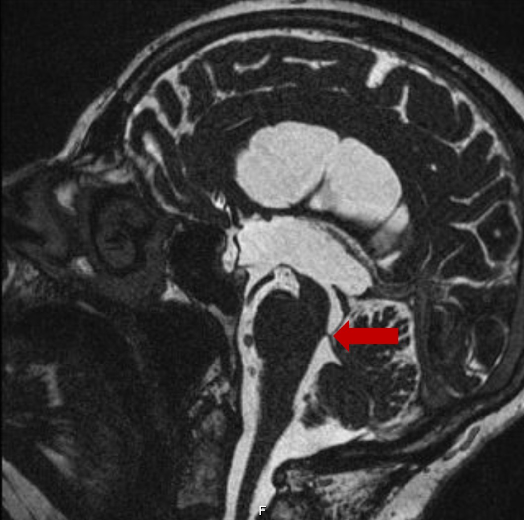
Cranial magnetic resonance (sagittal cut): presence of supratentorial ventriculomegaly, with a hyperintense halo surrounding the ventricular cavities, visualized in FLAIR, corresponding to gliosis, with no aspects suggesting the presence of transependymal exudation. These aspects are due to the presence of an obstacle (membrane) for drainage of the CSF at the level of the Sylvius aqueduct. Turbulence of the CSF flow is observed in the ventricles.
BIBLIOGRAFIA
1. Mentrasti G, Scortichini L, Torniai M, Giampieri R, Morgese F, Rinaldi S, Berardi R. Syndrome of Inappropriate Antidiuretic Hormone Secretion (SIADH): Optimal Management. Ther Clin Risk Manag. 2020;16:663-72. doi: 10.2147/TCRM.S206066.
2. Grant P, Ayuk J, Bouloux PM, Cohen M, Cranston I, Murray RD, Rees A, Thatcher N, Grossman A. The diagnosis and management of inpatient hyponatraemia and SIADH. Eur J Clin Invest. 2015;45(8):888-94. doi: 10.1111/eci.12465.
3. Yoshino M, Yoshimi Y, Taniguchi M, Nakamura S, Ikeda T. Syndrome of inappropriate secretion of antidiuretic hormone associated with idiopathic normal pressure hydrocephalus. Intern Med. 1999;38(3):290-2. doi: 10.2169/internalmedicine.38.290.
4. Kumar S, Bhayani P, Hathi D, Bhagwati J. Hyponatremia initial presenting feature of normal pressure hydrocephalus in elderly patient: a rare case report. JGG 2018;66:156-7.
5. Oliveira LM, Nitrini R, Román GC. Normal-pressure hydrocephalus: A critical review. Dement Neuropsychol. 2019;1Jul-Sep;13(3):361. doi: 10.1590/1980-57642018dn13-020001.
6. Hurley RA, Bradley WG Jr, Latifi HT, Taber KH. Normal pressure hydrocephalus: significance of MRI in a potentially treatable dementia. J Neuropsychiatry Clin Neurosci. 1999;11(3):297-300. doi: 10.1176/jnp.11.3.297.
7. Daou B, Klinge P, Tjoumakaris S, Rosenwasser RH, Jabbour P. Revisiting secondary normal pressure hydrocephalus: does it exist? A review. Neurosurg Focus. 2016;41(3):E6. doi: 10.3171/2016.6.FOCUS16189.
8. Sahuquillo J, Rubio E, Codina A et al. Reappraisal of the intracranial pressure and cerebrospinal fluid dynamics in patients with the so-called Normal pressure hydrocephalus syndrome. Acta neurochir. 1991;112:5061. doi:10.1007/BF01402454.
9. Meyer JS, Yasuhisa K, Tanahashi N, Tachibana H, Kandula P, Cech DA et al. Evaluation of treatment of normal-pressure hydrocephalus. Journal of Neurosurgery. 1985;62 513-21.
10. Lee JH, Park DH, Back DB, Lee JY, Lee CI, Park KJ et al. Comparison of cerebrospinal fluid biomarkers between idiopathic normal pressure hydrocephalus and subarachnoid hemorrhage-induced chronic hydrocephalus: a pilot study. Med Sci Monit. 2012;18(12):PR19-25. doi: 10.12659/msm.883586.
11. Tullberg M, Hultin L, Ekholm S, Månsson JE, Fredman P, Wikkelsø C. White matter changes in normal pressure hydrocephalus and Binswanger disease: specificity, predictive value and correlations to axonal degeneration and demyelination. Acta Neurol Scand. 2002;105(6):417-26. doi: 10.1034/j.1600-0404.2002.01189.x.
12. Rado M, Oreković D, Klarica M. The role of mesencephalic aqueduct obstruction in hydrocephalus development: a case report. Croat Med J. 2021;62(4):411-419. doi: 10.3325/cmj.2021.62.411.
13. Peterson DT, Marshall WH. Letter: Polydipsia and inappropriate secretion of antidiuretic hormone associated with hydrocephalus. Ann Intern Med. 1975;83(5):675-6. doi: 10.7326/0003-4819-83-5-675_2.
14. Yoshino M, Yoshimi Y, Taniguchi M, Nakamura S, Ikeda T. Syndrome of inappropriate secretion of antidiuretic hormone associated with idiopathic normal pressure hydrocephalus. Intern Med. 1999;38(3):290-2. doi:10.2169/internalmedicine.38.290.
15. Jung KH, Chu K, Jeong SW, Hong YH, Park KI, Roh JK. Idiopathic normal pressure hydrocephalus predominantly with prolonged fever and hyponatremia. Neurology. 2003;61(4):554-6. doi: 10.1212/01.wnl.0000078196.

