A 99-year-old male was admitted to Emergency Department due to dyspnoea. As part of the examination an X-ray was performed, showing pulmonary consolidation, pleural effusion, and numerous muscular rice grain calcifications (Fig.s 1 and 2). The patient was admitted into General Medicine ward with the diagnose of acute congestive heart failure due to community-acquired pneumonia. Neurologic symptoms and muscular pain were absent, and he referred pig domestic breeding when younger. These imaging findings and epidemiology history is highly suggestive of cysticercosis.1 Patient was discharged after thirteen days in his basal status without any specific treatment to cysticercosis.
Cysticercosis is caused by pork tapeworm Taenia solium, in the larval stage (metacestode). The prevalence of cysticercosis is heterogeneous but it is higher in rural areas. This occurs because of pig breeding and due to worse sanitary conditions.2 After the ingestion of Taenia solium eggs, embryos (oncospheres) hatch in the small intestine, invade its wall and disseminate hematogenously to brain (neurocysticercosis, which can lead to neurologic symptoms) and/or extraneural sites (striated muscle, subcutaneous tissue and liver being the most common).1 Stages of cysticercosis include an initial (viable) phase, a degenerating (enhancing) phase and a nonviable (calcified granulomas) phase which may persists for many years. Intramuscular cysts often undergo calcification and may be detected radiographically as disseminated rice grain calcifications.3 Many cases of cysticercosis are asymptomatic and they are identified incidentally via radiographic imaging performed for other reasons. Specific therapy is not needed in patients at the calcified staged if they are asymptomatic.2,4 Eradication of cysticercosis would require better sanitary conditions to avoid human infection by tapeworm due to contaminated pork consumption.5
Figura I
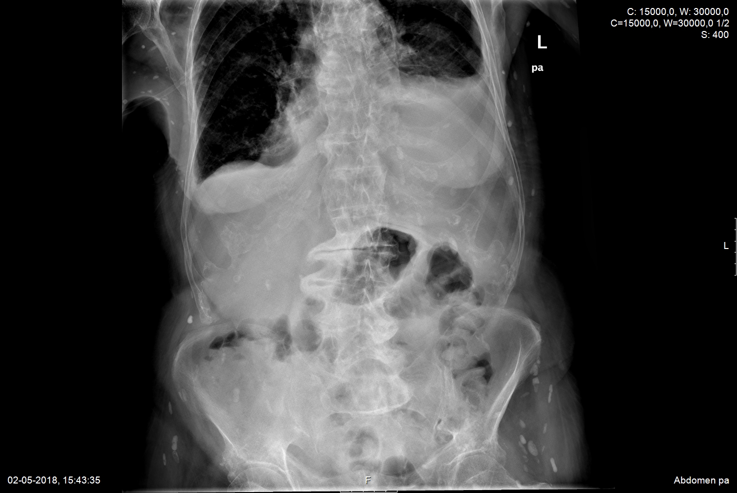
Abdominal X-ray showing typical fusiform, rice grain shaped calcifications, with orientation along the muscle plain.
Figura II
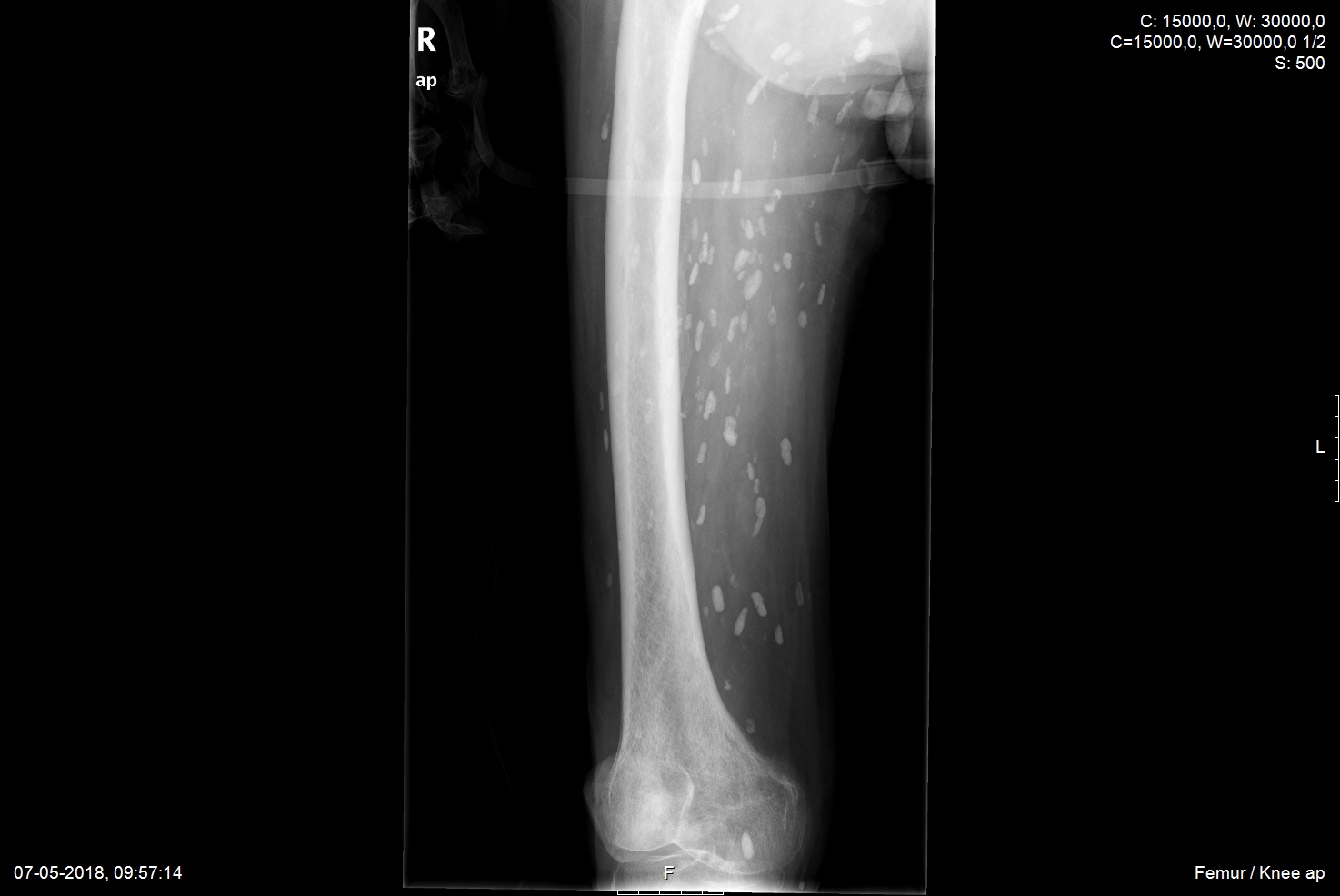
Femur X-ray showing typical fusiform, rice grain shaped calcifications, with orientation along the muscle plain.
BIBLIOGRAFIA
1. Del Brutto OH, Rajshekhar V, White AC Jr, et al. Proposed diagnostic criteria for neurocysticercosis. Neurology. 2001;57(2):177-183. doi:10.1212/wnl.57.2.177
2. White AC Jr, Coyle CM, Rajshekhar V, et al. Diagnosis and Treatment of Neurocysticercosis: 2017 Clinical Practice Guidelines by the Infectious Diseases Society of America (IDSA) and the American Society of Tropical Medicine and Hygiene (ASTMH). Clin Infect Dis. 2018;66(8):e49-e75. doi:10.1093/cid/cix1084
3. Bustos JA, Garcia HH, Dorregaray R, et al. Detection of muscle calcifications by thigh CT scan in neurocysticercosis patients. Trans R Soc Trop Med Hyg. 2005;99(10):775-779. doi:10.1016/j.trstmh.2005.04.011
4. Custódio M, Sequeira C, Marques A. Soft Tissue Cysticercosis: Rare Image in the Developed World. Acta Med Port. 2019;32(5):407. doi:10.20344/amp.11112
5. Garcia HH, Gonzalez AE, Tsang VC, et al. Elimination of Taenia solium Transmission in Northern Peru. N Engl J Med. 2016;374(24):2335-2344. doi:10.1056/NEJMoa1515520

